

Enhancing resolution and speed: How acousto-optic components are transforming confocal microscopy
G&H's Vice President of Life Sciences, Lars Sandström, reviews the use of acousto-optic components in confocal microscopy and options to overcome limitations in imaging depth, resolution, and speed in the recently published article of the January/February issue of BioPhotonics magazine.
In his article, "Acousto-Optic Components Overcome Limitations in Confocal Microscopy," Lars Sandström discussed how the use of acousto-optic deflectors (AODs) in confocal microscopy can address some of the limitations of traditional confocal and multiphoton microscopy techniques.
Confocal microscopy uses a continuous wave laser beam that is focused into a small (micron-wide) beam waist inside a tissue sample containing a fluorescent dye or protein. The resultant fluorescence passes back through the microscope objective and is then focused on a pinhole aperture located in front of a high-gain photodetector. This confocal aperture blocks any light that does not originate from the xyz location of the laser beam waist. By scanning the beam waist and/or moving the sample, a horizontal or vertical image slice or even an entire image cube can be acquired, with fluorescence captured at multiple depths.


Multiphoton microscopy is a related technique in which an ultrafast laser is focused using high-numerical-aperture optics. The laser wavelength is set to twice the wavelength needed for conventional excitation of the target fluorophore. At the beam waist, and only at the beam waist, the focused peak intensity exceeds the threshold for two-photon excitation. This provides inherent 3D resolution and eliminates the need for the lossy confocal aperture.
However, both of these techniques are negatively affected by practical trade-offs in imaging, such as the capability to image deeper within tissue at the frame rates necessary to capture metabolic processes. In addition, the resolution can be negatively affected by optical aberrations due to the microscope optics or, more insidiously, by the optical properties of the sample tissue itself.
Sandström explains that the use of acousto-optic deflectors (AODs) in confocal microscopy can help to address these limitations by scanning the laser using acousto-optic deflectors instead of galvanometers. In an acousto-optic configuration, an acoustic wave is applied to some type of optically transparent material, such as a crystal, that causes a change in the refractive index of the material. This change in refractive index causes the light passing through the material to be deflected. By applying a time-varying acoustic wave, the angle of deflection can be rapidly changed, allowing for fast and precise scanning of the laser beam.

AODs can be used to scan the laser beam in both the x and y axes, including arbitrary path of interest, providing a high-speed, high-resolution scan or of the sample. This can significantly improve the imaging depth and speed of confocal and multiphoton microscopy, making it possible to capture metabolic processes and other dynamic processes in real-time. Additionally, AODs can be used to modulate the intensity of the laser beam, making it possible to perform blanking during raster scanning, which can help to further improve resolution.
In summary, recent developments in acousto-optic and other active photonic components integrated into a microscope are beginning to address the limitations of confocal and multiphoton microscopy techniques, opening up new opportunities in life science research. The use of acousto-optic deflectors (AODs) in confocal microscopy has been found to be a promising solution for addressing limitations in imaging depth, resolution, and speed. This can significantly improve the ability to capture metabolic processes and other dynamic processes in real-time, providing new insights into the inner workings of living organisms.
The full article can be found in the January/February issue of BioPhotonics magazine or you can read the full publication on the Photonics.com website.
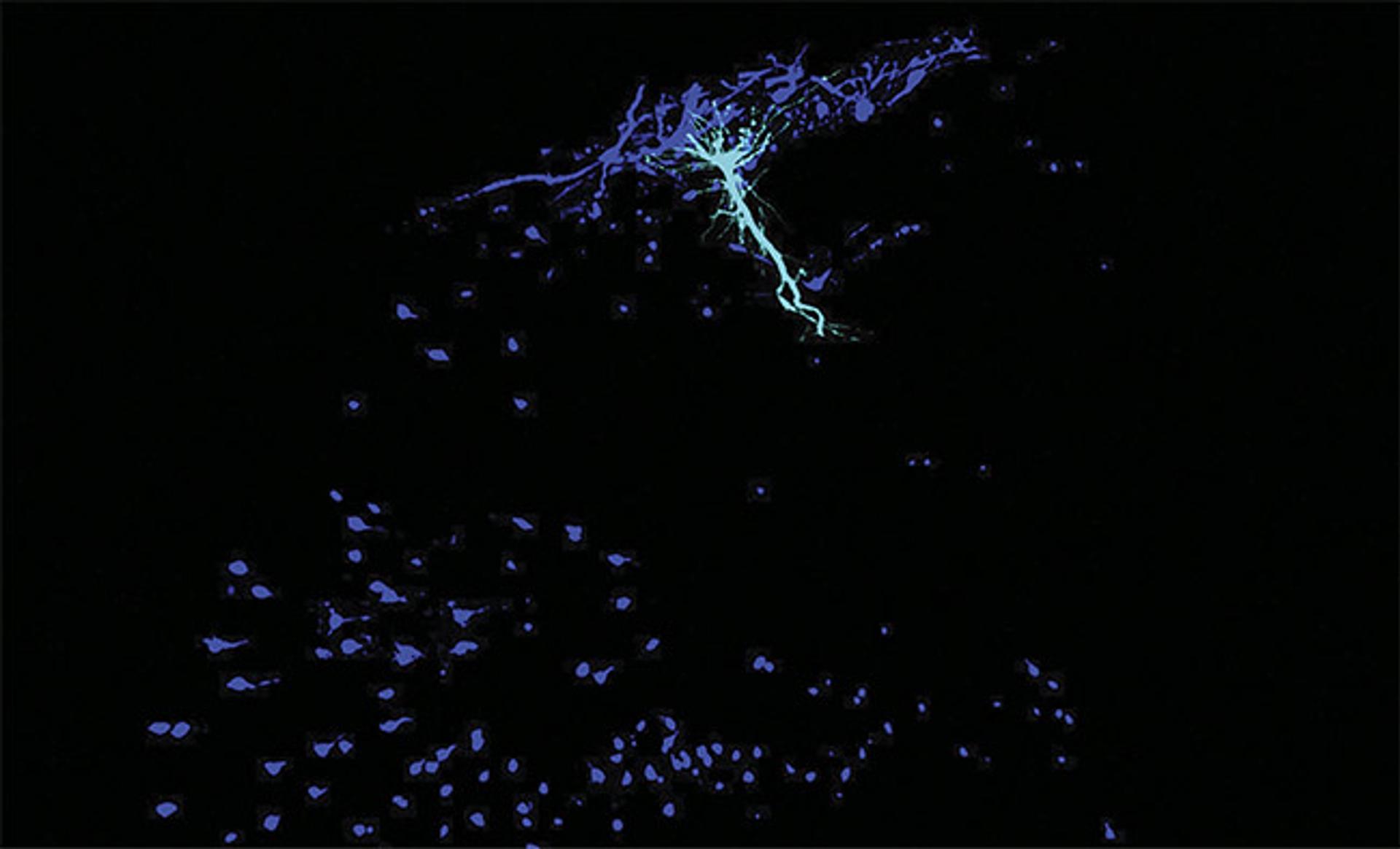
Image: A place cell (cyan) and its local circuit of interneurons (dark blue) in a mouse hippocampus. (Credit: Tristan Geiller / Losonczy lab / Columbia’s Zuckerman Institute)